At Casper & de Toledo, our Connecticut brain injury lawyer represents the victims and families throughout the state whose lives have been devastated due to an accident resulting in traumatic brain injury (TBI). Contact Casper & de Toledo today to learn more about brain injury diagnosis, what your diagnosis may mean for your future, and how we can help you fight for your rightful compensation.
Connecticut Traumatic Brain Injury Diagnosis
Any person who sustains an injury to his or her head can benefit from qualified representation. Scientific literature confirms that even people who experience whiplash from a car crash, truck accident, or any other type of accident can sustain a mild traumatic brain injury or worse. Every patient diagnosed with a concussion has sustained a traumatic brain injury of some magnitude. The key is to know and understand the signs and symptoms that indicate that whiplash or concussion has resulted in permanent damage to the brain. Sometimes the brain damage is microscopic, which means it exists on a cellular level and can produce damage to the brain’s neurons and/or axons. Similarly, a person can sustain an injury that causes intracranial bleeding, resulting in an epidural hematoma or a subdural hematoma without losing consciousness, without experiencing amnesia, and in some cases, without necessitating surgery. A head injury can also produce microscopic bleeding that exposes brain parenchymal tissue to blood products that can cause damage to the brain.
Many physicians throughout the country (including in Connecticut) do not focus on the subtle changes that indicate that a brain injury is present, mainly because in many cases, there is little, if any, treatment that can be provided to this type of patient. In other cases, certain physicians are really not trained to recognize subtle forms of brain injury. In emergency rooms, medical staff is often focused upon injuries that are life or limb-threatening, paying close attention to the patient’s airway, breathing, circulation, and more apparent disability. Mild TBI is frequently overlooked, and unless there is serious deterioration, most patients never return to the emergency room.
Here at Casper & de Toledo, we use our expertise to review symptoms that are often associated with brain injury, and we follow our clients as they “heal” to determine if the client, family members, or friends observe any changes in cognitive, physical, emotional, behavioral or sleep functioning. We use checklists to cover such items as reduced attention and concentration, memory problems, problems making decisions, depression, impaired judgment, dizziness/balance problems, blurred vision, ringing in ears (also known as tinnitus), alteration of the sense of taste, and/or smell, and many other indications. Often, these forms of deficits are only documented and recognized as a result of a specialized evaluation done by a neuropsychologist, an audiologist, a sleep specialist, or an otolaryngologist (ENT). In other cases, documentation of subtle forms of brain damage may be identified with specialized imaging techniques known as PET scans, SPECT scans, functional MRI evaluations (fMRI), or the latest technology available (including the use of a high-field MRI magnet, known as a 3.0 Tesla, together with Diffusion Tensor Imaging and volumetric assessment).
Diagnostic Tests and Traumatic Brain Injury
Microscopic gray and white matter injuries to the brain that occur often in the context of mild, complicated mild, and moderate traumatic brain injury will not be discernible on most diagnostic tests that are performed in an emergency department setting. For example, the gold standard for screening for acquired brain injury in an emergency room setting is the CT Scan. However, with the exception of the reliability of the CT Scan for documenting hemorrhage and/or skull fractures, the CT scan does not reveal much useful information about more subtle damage to brain structures. Likewise, the use of standard clinical MRI found in most community hospitals has limited utility in identifying subtle structural damage. Other tests may be relied upon to corroborate suspected brain injury, including:
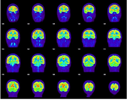

SPECT scan – assesses blood perfusion (or blood flow/spreading) in the brain.
Functional MRI (fMRI) – brain mapping that correlates structures within the brain with cognitive function.
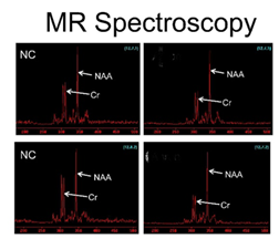

MR Spectroscopy – identifies metabolic changes in the brain tissue.
Electroencephalogram (EEG) – assesses the abnormal electrical activity in the brain, including seizure activity.
Diffusion Tensor Imaging (DTI) – measures the tendency for water to diffuse away from one location in the brain to another. Diffusion is constrained by numerous features of the local cellular environment in the brain, and it is therefore regarded as a measure of local tissue organization. One of the most frequently used DTI measures of tissue organization is termed “Fractional Anisotropy,” or FA. DTI analysis is performed by calculating the FA values for the patient and then comparing FA values at each point across the patient’s brain with FA values across the brains of healthy people.
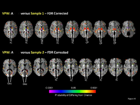

Brain Surface Analysis– Comparison of the cortical surface of the patient with the surfaces of healthy people using two measures. One is, strictly speaking, a measure of local indentations and protrusions at each millimeter-sized point across the surface of the brain. Because indentations and protrusions are caused by variations in underlying volumes of brain tissue, these measures are referred to as local volumes. The other measure is cortical thickness, or the thickness of the cortical mantle, the layer of gray matter that covers the surface of the brain and that primarily contains the nerve cell bodies, cellular appendages (dendrites), and connections (synapses) between neurons.
Perhaps the most exciting advances made in the last decade in the field of traumatic brain injury diagnosis are the advanced neuro-imaging techniques. Such studies are quite costly to administer and very costly to interpret. There are three very important concepts that underlie the ability of the lay public to understand these techniques.
It is commonly assumed that an MRI of a part of the body actually generates the images that are read by a radiologist or other specialist That is not so. The MRI equipment relies on a very strong magnet to acquire quantitative data (0s & 1s) that is used by different software programs to measure tissue structure. The quantitative data is then processed through the software that in turn generates different image formats depending upon the software used. The MRI settings must be established for a scan to conform with the data required for each program. In brain injury, microscopic structures are of critical importance, and therefore the strength of the MRI magnet is also critically significant, as many brain structures cannot be examined properly with the conventional MRI 1.5 Tesla magnet available in many locations.
The data collected from imaging studies and processed using imaging post-processing software can be compared to comparable data collected from scientifically selected controls that are the basis for the “normative data.” In this fashion, mathematicians, statisticians, and imaging experts can provide an analysis as to the extent that the patient’s quantitative data is different than the data available from the normative sample.
It is not the purpose of this summary to provide an exhaustive summary of the software options available for analyzing brain structures. However, two of the most useful computer sequences for assessing the structural integrity of the very small components of the brain are 1) Diffusion Tensor Imaging (“DTI”) that assesses the integrity of the brain’s white matter fibers; and 2) Volumetric Analysis that examines the brain volume for specific structures or brain segments.
White matter tracts (as distinct from the gray matter) can be analogized to the “wires” connecting the components or regions of the brain. Those fibers are known as “white matter” because they are covered by white myelin sheaths and are made up of different cellular than the gray matter. White matter fiber tracts are particularly susceptible to damage as a result of the trauma, and that damage can occur as a result of the partial or complete severing of the tract (“axotomy”) or can be damaged as a result of the stretching, twisting or compression of the fiber. The most common areas in the brain’s white matter to be impacted by trauma involved the gray/white matter junction. This area is particularly susceptible to injury because of the relative strength of the different tissue involved. The same biomechanical forces that are applied to the gray and white matter will more likely impact the white matter compared to the more resistant gray matter. The most useful tool for quantifying the structural integrity of white matter fiber tracts is the MRI computer sequence known as Diffusion Tensor Imaging (“DTI”). DTI measures the tendency for water to diffuse away from one location in the brain to another. Diffusion is constrained by numerous features of the local cellular environment in the brain, and it is therefore regarded as a measure of local tissue organization. One of the most frequently used DTI measures of tissue organization is termed Fractional Anisotropy, or FA. To assess the integrity of the fiber tracts, the fibers in a patient with an acquired brain injury are assessed by computing the FA values as previously described, and then the FA values at each point across the patient’s brain are compared with FA values across the brains of healthy people comprising a normative sample.
While the availability of obtaining diffusion tensor imaging for concurrent clinical and forensic purposes is limited, the technology is under widespread use in research and for some clinical purposes, including corroboration of probable brain injury. In litigation, there is documentation that DTI has been deemed admissible at trial in sixty-three cases and has only been precluded on three documented occasions. We believe that there are four primary reasons why this evidence is so controversial:
1) The injured party will rarely if ever, have baseline DTI imaging.
2) An image is powerful evidence (“A picture is worth a thousand words.”)
3) The findings consistent with white matter damage may possibly be related to other causes.
4) The technology is not diagnostic.
The inability of the study to confer a diagnostic conclusion should not bar the evidence that is based upon FDA-approved scientific technology and methodologies that have been the subject of peer review. The evidence is relevant in personal injury litigation because it tends to make the probability of brain injury more (or less) likely. When analyzed in the context of the locations of the white matter damage, and in the absence of co-morbid contributing causes, the evidence helps answer the question: Is it more likely than not that there was a brain injury? The clinician can reach a conclusion using the traditional differential process.
Two very useful scientific papers on the subject of diffusion tensor imaging in the context of so-called mild traumatic brain injury are: Shenton ME, Hamoda HM, Schneiderman JS, et al. “A review of magnetic resonance imaging and diffusion tensor imaging findings in mild traumatic brain injury”. Brain Imaging and Behav. 2012;6(2):137-192. doi:10.1007/s11682-012-9156-5 and Hulkower MB, Poliak DB, Rosenbaum SB, Zimmerman ME, Lipton ML. “A decade of DTI in traumatic brain injury: 10 years and 100 articles later”. ANJR Am J Neuroradiol. 2013;34(11):2064-2074. DOI: 10-3174/ajnr.A3395. The importance of diffusion tensor imaging for identifying structural connectivity changes in the brain in acute and chronic stages of traumatic brain injury and predicting a patient’s long-term outcome has been recognized in leading medical journals, including Neurology, which published “Longitudinal changes in structural connectivity of traumatic axonal injury” in 2011. Wang JY, Bakhadirov K, Abdi H, et al. Longitudinal changes of structural connectivity in traumatic axonal injury. Neurology. 2011;77(9):818-826. DOI: 10.1212/WNL.0b013e31822c61d7. More recent papers published in neurology journals have supported the contention that the physiological changes within the brain continue after clinical recovery. Brett BL, Wu YC, Mustafi SM, et al. “The Association between Persistent White-Matter Abnormalities and Repeated /Injury after Sport-Related Concussion”. Front Neurol. 2020;10:1345. Published 2020 Jan 21. doi:10.3389/fneur.2019.01345.
Volumetric analysis of the brain is capable of examining at least three levels of brain structures. First, brain imaging experts can assess the overall volume of the brain. Second, an analysis of the cortical surface of the brain comparing it to the normative data measuring the same regions in a normative sample can be performed. These calculations are assessments of local indentations and protrusions at each millimeter-sized point across the surface of the brain. Because indentations and protrusions are caused by variations in underlying volumes of brain tissue, these measures are often referred to as local volumes. The third measure is cortical thickness, or the thickness of the cortical mantle, the layer of gray matter that covers the surface of the brain and that contains primarily the nerve cell bodies, cellular appendages (dendrites), and connections (synapses) between neurons. All of these measurements can be useful for identifying discrepancies between the structural anatomy of the patient’s brain in relation to a normative sample, as well as based upon an absence of symmetry in an organ known to have symmetrical features.
Below is an example of a portion of a DTI study that reflects a normative image of the corpus callosum on the left and the damaged corpus callosum on the right.
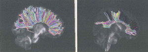

The following illustrations are from a case involving a 9-year-old boy who suffered a “so-called” mild traumatic brain injury in a car accident. Of course, when you look at the damage to his brain, one would seriously question the label “mild.”
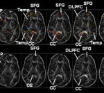

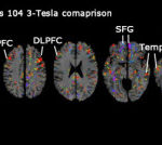

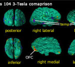

The common misperception about advanced neuroimaging is that it is used as a biomarker to diagnose a traumatic brain injury. It is not a diagnostic tool. Rather, it is a tool that can identify structural brain damage that may be considered consistent or inconsistent with brain injury. The opinion must be arrived at through a differential clinical analysis, where a physician arrives at the most probable diagnosis based upon a panoply of factors.
To date, detractors of the use of DTI as a component of the differential diagnostic process for mild traumatic brain injury have prevailed in precluding DTI evidence in only 0.5% of challenges. The challengers fear that the science is proof of structural irregularities that otherwise could only be confirmed on autopsy, clearly not an option for living human beings.
The Role of Neuropsychology in the Evaluation of Head Injuries
Complete evaluation of the consequences of traumatic brain injury most often requires a neuropsychological evaluation. Neuropsychology is a specialized field within the field of psychology. A neuropsychologist administers a battery of standardized tests of cognitive function, personality, behavior, and effort. Most of the tests have been thoroughly researched and normed so that an opinion can be developed as to whether or not the patient has experienced any decrease in the type of functions that are sensitive to a brain injury. But some of the instruments have never been normed or standardized on a traumatic brain injury population, and the interpretation of the results can be unfairly manipulated. Results from some of the instruments can be easily manipulated.
A good and honest neuropsychologist should engage in the differential diagnostic process. A differential diagnosis is a process by which a health care provider considers the patient’s prior history, the history of the present illness or condition, the signs and presenting symptoms, and other available data, and through the process of elimination, determine the most probable diagnosis that fits the presentation. In medicine, in a differential diagnosis, a physician would continue to consider life and limb-threatening conditions until able to rule out such conditions. A neuropsychologist will consider whether the presenting signs and symptoms might be consistent with 1) a medical condition; 2) psychopathology; and 3) traumatic brain injury. With regard to a general medical condition, there are inventories that might be given to an examinee that are intended to highlight responses suggestive of such conditions. The MMPI-2 or the MMPI-2-RF were intended to identify psychopathology and often reveal elevated scales that may be entirely consistent with a traumatic brain injury population but might also suggest that someone without a brain injury may be over-reporting symptoms, may not be using adequate effort, may lack in motivation, or maybe engaging in impression management. In the case of the MMPI-2 and the MMPI-2-RF, there has been much scientific criticism because some psychologists attempt to use the instrument to identify malingerers – patients who are allegedly intentionally faking their condition. The American Psychological Association has expressed severe reservations about the use of the Fake Bad Scale (“FBS”), also known as the Symptom Validity Scale, and several courts around the country have excluded the use of the FBS because it is scientifically unreliable and is inherently biased against patients suffering from traumatic brain injury, and women in particular. Stewart Casper was interviewed by the Connecticut Law Tribune concerning the Fake Bad Scale in September 2008. The Fake Bad Scale is comprised of 43 of the 567 items contained in the MMPI-2. Each item is answered “True” or “False.” If a patient’s responses to the FBS items results in a score of 26 or higher, the patient is identified as a malingerer. Yet the items in the FBS consist of complaints and symptoms common to people who have suffered a serious injury, including acquired brain injury. Certainly, in multi-trauma cases, there is good reason to look at some scale elevations with skepticism.
Other tests administered by neuropsychologists, or perhaps even some neuropsychiatrists, are used to measure effort. People taking these tests must always use their best efforts – always do their best and always tell the truth. The way these assessments are constructed, administered, and scored results in “red flags” being raised by unusual results. While there is no test that can actually diagnose someone as a faker or an exaggerator, the “red flag” is just enough to ruin a case. And there are neuropsychologists ready and willing to exploit unusual test and personality inventory results for his or her secondary gain. Therefore, it is important for an accident lawyer to be capable of recognizing the limitations of tests that can easily be manipulated to mischaracterize the performance of a brain-injured patient.
Interestingly, there is an increasing recognition by experts and trial judges that to identify a test that contains the pejorative word “malingering” (as in “Test of Memory Malingering” (“TOMM”)) can be unfairly prejudicial to a litigant in front of a jury. The test itself does not diagnose a condition or a level of effort. Rather, a performance that does equal or exceeds the cut score raises a red flag about possible poor effort. Standing alone, a substandard score should not result in an unflattering diagnosis. Tests do not make a diagnosis; clinicians make the diagnosis. And it would be difficult to justify a “poor effort” or “malingering” diagnosis taking into account performance on all effort measured. Whether a party or witness is telling the truth is solely within the province of the jury to decide.
Your Concussion Is a Brain Injury
Health care professionals, including academics, have been at odds for years about the methodologies for classifying concussions. Needless to say, uniformity in descriptors has been elusive. In 1966, the Congress of Neurological Surgeons defined concussion to be a “clinical syndrome characterized by immediate and transient posttraumatic impairment of neural function, such as alteration of consciousness, disturbance of vision, equilibrium, etc. due to brain stem involvement.” Other researchers have defined concussion by distinguishing between a “mild concussion,” a “moderate concussion” and a “severe concussion,” relating severity to the extent of loss of consciousness (“LOC”) with mild equating to “no LOC;” moderate equating to brief LOC plus retrograde amnesia, and severe equating to LOC for five minutes or more. Another researcher (Cantu 1986) developed definitions for concussion differentiating between Grade 1, 2, and 3. Grade 1 would be a concussion with no LOC and less than 30 minutes of post-traumatic amnesia; Grade 2 would be LOC for less than five minutes or post-traumatic amnesia lasting longer than 30 minutes but less than 24 hours; and Grade 3 concussion would refer to injuries accompanied by a LOC of greater than 30 minutes or post-traumatic amnesia lasting longer than 24 hours. Cantu later revised his concussion criteria in 2001 to combine Grades with a duration of symptoms of Post Concussion Syndrome (“PCS”), using the signs or symptoms of PCS that can be verified by neuropsychological tests. Another widely used guideline for rating concussions is the Practice Parameters for Concussion Severity adopted by the American Academy of Neurology (“AAN”). The AAN likewise uses three grades for concussion ranking severity as follows: Grade 1 (mild): “Transient confusion; symptoms or mental status abnormalities on examination resolve in less than 15 minutes and no LOC”.; Grade 2 (moderate): “Transient confusion; symptoms or mental status abnormalities on examination last longer than 15 minutes and no LOC”; Grade 3 (severe): “Any LOC either brief (seconds) or prolonged (minutes).”
Often, accident victims experience a direct blow to the head or even a severe shaking of the head, which can occur in a rear-end collision and whiplash type of injury. This is also known as an acceleration-deceleration injury. The common feature of both types of injuries is the sudden movement of the skull that causes the brain, with a gelatinous-like consistency, to bounce around inside the skull. The inside of the skull contains a number of rough and jagged bony ridges that can cause injury to the brain. Furthermore, the forces, including rotational forces, can cause damage within the brain.
More often than not, the damage inflicted in these types of events cannot be seen on CT scans or standard MRI. Yet the patient’s history, combined with the constellation of symptoms, leaves little doubt that blows to the head, often dismissed as simple concussions are in fact brain injuries that may have permanent adverse consequences. Moreover, very often a patient is discharged from a hospital emergency room after having a CT scan and being evaluated. A discharge does not mean there is no brain injury. One should not get a false sense of security, because the persistence of symptoms should warrant a visit to a neurologist or doctor of physical medicine and rehabilitation that specializes in head injury.
Find Help from a Skilled Brain Injury Attorney
No other law firm in the state of Connecticut provides you with the type of background we offer in dealing with brain injury cases. Partner Stewart Casper and his Connecticut brain injury team are members of a select group of lawyers throughout the nation who have attended advanced seminars on the specifics in the areas of brain imaging, neuropsychology, life care planning, and retention of experts. Mr. Casper has attended these advanced educational programs for lawyers, and he has also taught at several of the programs. Among the programs he has attended is “Advanced Techniques in Neuroimaging,” which included subjects on CT Scans, Magnetic Resonance Imaging, MR Spectroscopy, Functional MRI, Diffusion Tensor Imaging, PET Scans, and SPECT Scans. He has taught classes throughout the United States on brain injury subjects including:
- “Make the Defense Talk about the Science”
- “Cross-Examination of the Defense Neurologist”
- “Mining the Traumatic Brain Injury Literature”
- “Broken Connections & Brain Networks”
- “Proving and Winning the Traumatic Brain Injury Case”
- “The Selection and Use of Experts in the Traumatic Brain Injury Case”
- “Proving the Injury in the Traumatic Brain Injury Case”
- “Cross-Examination of the Defense Expert in a Traumatic Brain Injury Case”
- “Misclassification of Mild Traumatic Brain Injury”
- “Debunking the Myths of TBI”
- “Strategies for Diffusion Tensor Imaging & Volumetric Analysis in Pediatric TBI Litigation”
- “Proving Traumatic Brain Injury with Demonstrative Evidence”
- “The Progressive Injury Cascade of Traumatic Brain Injury”
Contact a Connecticut Brain Injury Lawyer from Casper & de Toledo
Our traumatic brain injury lawyers help residents in Bethel, Bridgeport, Brookfield, Danbury, Darien, Easton, Fairfield, Greenwich, Monroe, New Canaan, New Fairfield, Newtown, Norwalk, Redding, Ridgefield, Shelton, Stamford, Stratford, Trumbull, Weston, Westport & Wilton and other cities and towns of Connecticut to help in their pursuit of compensation from the parties who are responsible for their traumatic brain injuries. Contact Casper & de Toledo today to schedule your initial consultation with our firm.

